Summary of New Imaging Tech Creates 3D Maps of Uterine Contractions During Labor in Real-Time:
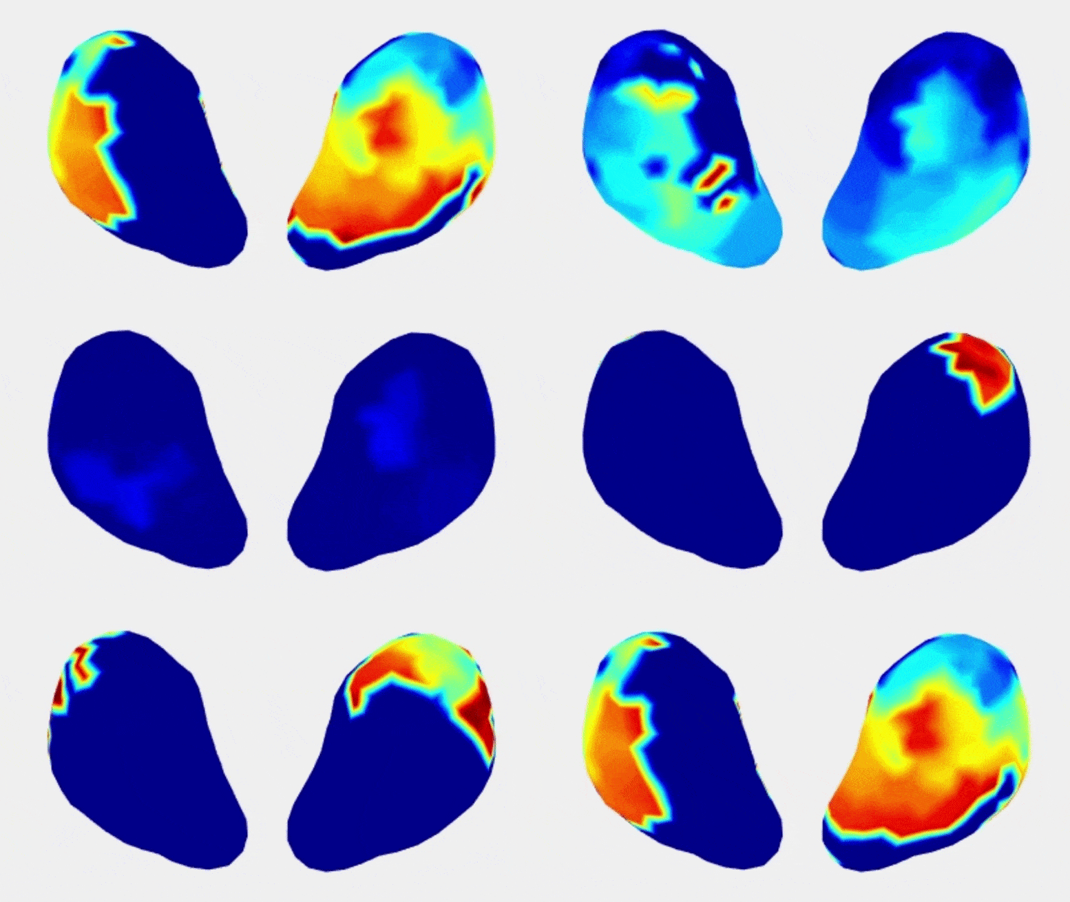
Researchers at Washington University School of Medicine have developed a new imaging technique that can create 3D maps showing the magnitude and distribution of uterine contractions during labor in real-time. Called electromyometrial imaging (EMMI), the technology can be used to identify and monitor complications in high-risk pregnancies and to prevent preterm birth, which affects 10% of pregnancies globally. The researchers are also working on a low-cost EMMI system that could be used in resource-poor areas with less expensive and more portable ultrasound imaging. The study was published in the journal Nature Communications.
*****
Washington University School of Medicine in St. Louis researchers have developed a new imaging technique that could help detect problems like preterm labor or labor arrest. The noninvasive technology can produce real-time 3D maps of uterine contractions, shedding light on early contractions that lead to preterm birth and identifying ways to slow or stop these contractions. The technology, called electromyometrial imaging (EMMI), was developed based on imaging methods used for the heart, but with greater detail, allowing for a four-dimensional mapping of labor contractions. The technology could be used to optimize pharmaceutical treatment, monitor uterine contractions, prevent postpartum hemorrhage, provide non-pharmaceutical treatment options, and research other uterine-related conditions.
The study included 10 participants in labor through childbirth, and researchers found that uterine contractions are less predictable and consistent than heart contractions. Consecutive labor contractions can differ in the initiating region and direction of progression, and there are no consistent areas of the uterus in which contractions begin, indicating the uterine pacemaker is not anatomically fixed, like the heart. Patients who had not given birth before had longer contractions with more variation, while those who previously had given birth had more efficient and productive contractions.
The next step for researchers is to measure normal uterine contractions to better understand whether a contraction is productive and leading towards birth. The research team has received a grant from the National Institutes of Health to characterize what contractions during normal labor look like, aiming to create a normal baseline atlas. The new imaging technology could help make labor and childbirth safer, especially in resource-poor areas. To increase accessibility, the team is developing a low-cost EMMI system using printed disposable electrodes and a wireless transmitter. The team also hopes to broaden research to other uterine-related conditions outside of pregnancy, such as painful menstruation and endometriosis.
In conclusion, the new technology developed by researchers at Washington University School of Medicine in St. Louis could revolutionize the way uterine contractions are monitored by providing real-time 3D maps of the contractions in full detail. The technology would help improve the accuracy with which uterine contractions are measured and detected, which could ultimately lead to better care for pregnant women with high-risk pregnancies, the prevention of preterm birth, and a reduction in the number of C-sections, which can increase the risk of birth injuries or death. In addition, the technology could be used to advance research into other uterine-related conditions like painful menstruation and endometriosis.


Comments are closed