Summary of A Game-Changer for Personalized Medicine:
Researchers from the Georgia Institute of Technology and Emory University have developed an intracellular toolkit for studying organelle diversity and communication within stem cells, which includes rapid subcellular proteomic imaging and multiplexed immunofluorescence techniques. Using this toolkit, they could create maps of organelle organization and determine which cell types would best treat various diseases, leading to improved personalized cell therapies by identifying and isolating distinct stem cell subsets. The researchers also combined machine learning and spatial transcriptomics to analyze the spatial organization of multiple neighboring RNA molecules in single cells, providing insight into the importance of RNA-RNA proximity for accurate cell type classification.
*****
Researchers Develop Intracellular Toolkit to Study Organelle Diversity and Communication within Stem Cells
Georgia Institute of Technology and Emory University researchers have developed an intracellular toolkit to study organelle diversity and communication within stem cells. This toolkit includes rapid subcellular proteomic imaging and multiplexed immunofluorescence techniques, which allowed the researchers to create maps of organelle organization and determine which cell types would best treat various diseases. The researchers believe this toolkit will lead to more accurate cell type classification and improved personalized cell therapies by identifying and isolating distinct stem cell subsets.
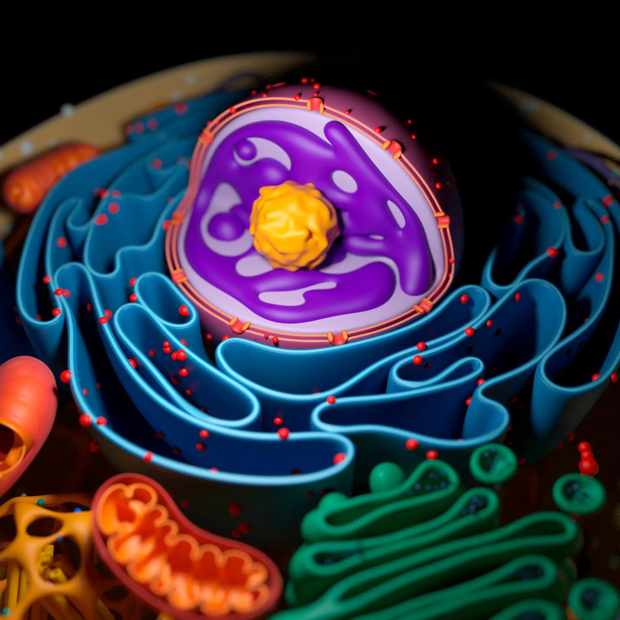
Organelles are bits and pieces of RNA and protein within a cell that play essential roles in human health and disease, such as maintaining homeostasis, regulating growth and aging, and generating energy. Organelle diversity in cells exists between cell types and between individual cells. Studying these differences helps researchers better understand cell function, leading to improved therapeutics to treat various diseases.
Creating the Subcellular Omics Toolkit
The first study looked at mesenchymal stem cells (MSCs) that have historically offered promising treatments for repairing defective cells or modulating the immune response in patients. In a series of experiments, the researchers created a data-driven, single-cell approach through rapid subcellular proteomic imaging that enabled personalized stem cell therapeutics.
The researchers then implemented a rapid multiplexed immunofluorescence technique using antibodies targeting specific organelles. By fluorescing antibodies, they tracked wavelengths and signals to compile images of many different cells, creating maps. These maps enabled researchers to see the spatial organization of organelle contacts and geographical spread in similar cells to determine which cell types would best treat various diseases.
RNA-RNA Proximity Matters
In the following study, the researchers took the toolkit further by studying the spatial organization of multiple neighboring RNA molecules in single cells, which are essential to cellular function. The researchers evolved the tool by combining machine learning and spatial transcriptomics. They found that analyzing gene proximity variations for cell type classification was more accurate than analyzing gene expression only.
The experiment consisted of two parts: the development of computational methods and investigations at the lab bench. The researchers examined published datasets and an algorithm to group RNA molecules based on their physical location. This “nearest neighbor” algorithm helped determine gene groupings. On the bench, researchers then labeled RNA molecules with fluorescents to quickly locate them in single cells. They then uncovered many features from the distribution of RNA molecules, such as how genes are likely to be in similar subcellular locations.
Cell therapy requires many cells with highly similar phenotypes. If there are subtypes of unknown cells in therapeutic cells, researchers cannot predict the behavior of these cells once injected into patients. With these tools, more cells of the same type can be identified, and distinct stem cell subsets with uncommon gene programs can be isolated.
“We are expanding the toolkit for the subcellular spatial organization of molecules – a ‘Swiss Army Knife’ for the subcellular spatial omics field if you will,” said Ahmet F. Coskun, a Bernie Marcus Early Career professor in the Coulter Department of Biomedical Engineering at the Georgia Institute of Technology and Emory University. “The goal is to measure, quantify, and model multiple independent but also interrelated molecular events in each cell with multiple functionalities. The end purpose is to define a cell’s function that can achieve high energy, Lego-like modular gene neighborhood networks and diverse cellular decisions.”